


New Treatment for Macular holes
Recent approval of Jetrea (Ocriplasmin) to treat symptomatic VMA (Vitreo Macular Adhesion) has opened up a new era in non surgical management of macular holes. Previously only a hospital based surgery was able to reverse the process of macular hole development but now a new drug can be injected into the eye painlessly in the office and within a few weeks the process reverses back to more normal vision levels.... more
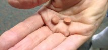
FDA Approves ASRS Leader's Argus� II Artificial Retina
On February 14, the FDA approved the Argus II artificial retina developed by ASRS Executive Committee and Board Member Mark S. Humayun, MD, PhD.
This breakthrough technology is the first ever to offer limited vision to patients with late-stage retinitis pigmentosa (RP).
Second Sight Medical Products (Sylmar, CA) manufactures the Argus II implant, which has 60 electrodes and a tiny camera mounted on eyeglasses to capture images.
The FDA approved Argus II for adults age 25 years or older with severe to profound RP. About 10,000 to 15,000 of the 100,000 Americans with RP will qualify for Argus II. Up to 4,000 patients a year can be treated with the device.
Macular degeneration and Aspirin?
Aspirin and Macular degeneration... is there an increase in the wet form of macular degeneration in those patients taking aspirin? The wet form develops quicker than the dry form. It may only be related to the fact that you are more likely to take aspirin if you have medical problems like stroke or heart attacks in the past and these conditions are more commonly associated with wet age related macular degeneration. ... more

Retinal Detachments & Tears
Overview:
The retina is a neurosensory tissue that lines the back inside wall of the eye, and is responsible for transferring incoming light into vision. When a tear or a detachment occurs, the retina is separated from the back wall of the eye, and thereby deprived of nutrition and blood. Without the vital nutrients, the retina will degenerate and result in vision loss.
Typically, retinal detachments occur in individuals over the age of 50, although blunt trauma or being nearsighted can cause a detachment in a younger individual. Other risk factors include diabetes, rubbing the eyes or recent cataract surgery.
At East Carolina Retina Consultants, Dr. Van Houten is well-versed in diagnosing and surgically treating cases of retinal detachment and can help you discover the most effective treatment plan for you.
Symptoms & Types:
Most tears occur in the peripheral retina, which will cause a loss in a portion of side vision initially after a detachment. Common symptoms of a retinal detachment are floaters or flashes in your field of vision. Floaters are strings or clumps of the liquid vitreous (from the center of your eye ball) that have aged and broken off; appearing as strings, globs or dots. Flashes indicate being able to see flashes of light or brief sparks at the edge of the vision field when moving your eyes or your head. Some also experience a sudden loss of vision in part of their visual field. Do not ignore these symptoms, but seek an exam from one of our professionals as soon as possible as retinal detachments are a serious concern that should not been taken lightly.
Unfortunately, some tears and breaks detach the retina completely without any warning symptoms. However, over 90% of retinal detachments can be repaired with a single surgical procedure. If multiple procedures are needed then an overall rate of sucessful retinal detachment repair reaches 95-98%. Note that sucess is defined as the reattachement of the retina, not what the vision will be. The vision will return as best it can depending on factors that are out side of physician control. They can incude: how long the retina was detached, did the central vision become detached, how much scar tissue did your body form and many more issues. As long as the retina remains attached, the vision will slowly improve over the coming years to the best of it's ability to recover.
The three types of retinal detachments are:
Rhegmatogenous Retinal Detachment: A result of a tear, during this type of detachment fluid from the center of the eyeball (called “liquid vitreous”) seeps into the back of the eye (through an open tear or break) and “lifts” the retina from the back of the eye.
Exudative Retinal Detachment: During this detachment, fluid called exudate leaks from blood vessels under the retina, causing the retina to detach. These can be caused by tumors or inflammatory disorders.
Traction Retinal Detachments: When this detachment occurs, fibro-vascular (scar) tissue within the vitreous cavity is pulling on the retina and causing the separation. Most often, these detachments are caused by proliferative diabetic retinopathy or history of significant bleeding in the eye.
Treatment:
It is ideal if a break in the retina can be discovered before a full detachment occurs. With a break or tear, a laser can be used to burn the tissue surrounding the break, which will then scar and heal through the retina. Another alternative is using a retinal cryoprobe to freeze the break, while offering the same scarring and healing effect of the laser. This is considered surgery as the anatomy of the eye is changed by the laser or cryo however no cutting or traditional surgery is done.
If a break has cause a complete detachment of the retina, then surgery will be needed.
There are 3 procedures for surgically treating a partial or total retinal detachment:
This procedure is ideal for early, uncomplicated rhegmatogenous retinal detachments with a single break located in the superior part of the retina.
After numbing the eye, the surgeon will inject a gas bubble into the middle of the eye, and use the bubble to assist in floating the detached retina back into position by moving the head accordingly. The bubble will help to seal the tear and flatten the retina down, sealing it to the eye wall. As long as the break is covered, the subretinal fluid will resolve in 1-2 days. The remaining tear is then treated either with a laser or a cryoprobe; creating scar tissue and healing the break. The patient will need to position for up to two weeks after treatment to hold the bubble in place closing the hole and allowing the retina to heal. The bubble will resolve with time (usually 4-6 weeks). No flying in an airplane while there is a bubble in your eye. There are usually post op drops to use on a schedule.
Scleral Buckling, up until about two decades ago was one of the only procedures to treat retinal detachments effectively and safely. It is a tried and sucessful technique that has stood the test of time. Ideal for a retinal detachment involving multiple breaks or tears in a patient that still has their natural lens in place. A scleral buckle is typically a piece of silicone implant (solid or sponge form), formed to act like a belt surrounding the middle part of the eye, that is used to close the retinal tears after they have been treated with a cryoprobe to speed healing. The buckle is sewn to the eye’s outer wall so that it changes the shape of the eye promoting closure of the retinal tears. It is hidden under the eye lids under a layer of conjunctiva (skin of the eye ball) and is a semi permanent addition to the eye. It can be removed later if it causes problems however that is rarely the case and would need another surgery to remove it. The procedure can typically be done with local or general anesthesia at the hospital.
Following the operation, most are able to resume normal levels of activity within several weeks.
A procedure first used over 30 years ago and continuously refined by modern medicine, vitrectomy surgery is used to treat traction retinal detachments (caused by diabetes) or more complicated rhegmatogenous detachments associated with vitreous traction or a vitreous hemorrhage.
Small incisions are made in the eye wall to give the surgeon better access to your retina. Then, the vitreous gel that fills your eyeball is removed as it contains the blood and scar tissue that is causing the problem. Without the gel, the surgeon can also better access your retina and uses a variety of instruments and techniques to seal up the tears. The gel is then replaced with a solution that mimics its composition. This procedure is also often done as a same day surgery with local or general anesthesia in the hospital. Post-operation, a specific head position may be required to keep a gas bubble pushing the retina into position while it heals. Silicone oil can also be used as a vitreous substitute to hold the retina in place but is used in more complicated cases or when the patient cannot position with a bubble. It will need another surgery at least 3 months later to remove the oil when the retina is stable.
Prevention:
Many retinal detachments are a result of older age and other medical problems, such as long-term diabetes. Have regular eye exams and physicals to rule out any vision problems and to manage medical problems like diabetes.
Even without hereditary risk factors, you should always wear eye protection when doing any activity where a small object could damage your eye (such as gardening, mowing the lawn, or sawing) or any activity where you could receive a blow to the eye (like basketball, baseball, shooting, etc).