


New Treatment for Macular holes
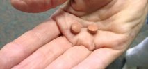
Recent approval of Jetrea (Ocriplasmin) to treat symptomatic VMA (Vitreo Macular Adhesion) has opened up a new era in non surgical management of macular holes. Previously only a hospital based surgery was able to reverse the process of macular hole development but now a new drug can be injected into the eye painlessly in the office and within a few weeks the process reverses back to more normal vision levels.... more
FDA Approves ASRS Leader's Argus� II Artificial Retina
On February 14, the FDA approved the Argus II artificial retina developed by ASRS Executive Committee and Board Member Mark S. Humayun, MD, PhD.
This breakthrough technology is the first ever to offer limited vision to patients with late-stage retinitis pigmentosa (RP).
Second Sight Medical Products (Sylmar, CA) manufactures the Argus II implant, which has 60 electrodes and a tiny camera mounted on eyeglasses to capture images.
The FDA approved Argus II for adults age 25 years or older with severe to profound RP. About 10,000 to 15,000 of the 100,000 Americans with RP will qualify for Argus II. Up to 4,000 patients a year can be treated with the device.
Macular degeneration and Aspirin?
Aspirin and Macular degeneration... is there an increase in the wet form of macular degeneration in those patients taking aspirin? The wet form develops quicker than the dry form. It may only be related to the fact that you are more likely to take aspirin if you have medical problems like stroke or heart attacks in the past and these conditions are more commonly associated with wet age related macular degeneration. ... more

Vitrectomy Procedures
Overview:
The Vitrectomy procedure was first used over thirty years ago to treat retinal detachments, and has been refined several times since then. A vitrectomy is most often used to treat tractional retinal detachments that typically result from proliferative diabetic retinopathy. These types of detachments occur when fibro-vascular tissue within the vitreous cavity pulls on the retina, causing it to separate from the eye wall. Other reasons include epiretinal membranes, macular holes, non clearing vitreous hemorrhages, and complicated retinal detachments.
Process:
If you and your physician at East Carolina Retina elects to perform a vitrectomy, you will have a surgical work-up approximately one week prior to surgery. The day of your procedure, you will be admitted to the hospital as an out patient, and can go home the same day. Over night stay is possible if you are coming from a long distance or surgery is late in the day as you need to be seen the first post op day. There are hotels close by for those that do not want to stay in the hospital overnight. General or local anesthesia can be used however general is preferred as it is easier on the patient and there is an excellent anesthesia department at Vident Hospital. During your vitrectomy, your surgeon will make small incisions into the wall of the eye to allow instruments to pass into the vitreous cavity (the middle of the eyeball). Your vitreous, or the fluid that is in the center of your eyeball, is then removed using a vitreous cutter. Delicate instruments are used to peel any scar tissue away from the retina. Once the vitreous, scar tissue or blood is out, your surgeon will use necessary techniques to re-attach your retina or finish the procedure.
Following surgery, if you have a gas bubble, you may be instructed to keep your head in a certain position to keep your retina attached while it heals. Your eye may be red, swollen and tender for several weeks; you will be provided with medications to reduce symptoms of pain, swelling and risk of infection. Be sure to follow all physician recommendations closely on the post op sheet that is given to you at the hospital, including activity guidelines and the placement of your eye patch (if required). Bring the post op sheet and the bag of eye medications to your post op visits each time so that they can be reviewed and changed as your eye heals.
Benefits:
- Can stabilize or return vision lost due to severe retinal problems.
- Can remove scar tissue, blood and/or debris that may be blurring the vision.
- Can reattach the retina and stabilise proliferative retinopathy.
Risks:
- Procedure carries similar risks of any invasive surgery, including: infection, bleeding, high eye pressure, retinal detachment, retinal tear, cataract formation, glaucoma, vitreous hemorrhage and development of scar tissue, loss of vision, loss of eye, pain.
- Vision may take months to improve, and may not ever recover completely depending on many factors.
- General anesthesia carries rare side effects such as: nausea, vomiting, heart problems, stroke, hiccupping and confusion. Death due to anesthesia is extremely rare.
- May need multiple procedure to correct the original or new problems.